Uncategorized

Interesting facts about Vitamin D:

Vitamin D insufficiency affects almost 50% of the population worldwide. Vitamin D was used in the pre-antibiotic era for the treatment of patients with tuberculosis (TB), when the ancient Greeks had first introduced “heliotherapy” (i.e., sunlight exposure) to treat TB. Vitamin D deficiency is linked with cardiovascular disease, anxiety, depression. Overview of Vitamin D Vitamin D is a fat-soluble vitamin. (Fat soluble vitamins are Vitamin A,D,E,K). Very few foods naturally contain vitamin D (fatty fish are the exception), so dermal synthesis is the major natural source of the vitamin.(From Skin) Vitamin D from the diet or dermal synthesis is biologically inactive and requires enzymatic conversion to active metabolites. Vitamin D is converted enzymatically in the liver to 25-hydroxyvitamin D (25[OH]D), the major circulating form of vitamin D, and then in the kidney to 1,25-dihydroxyvitamin D, the  active form of vitamin D. Vitamin D and its metabolites have a significant clinical role because of their interrelationship with calcium homeostasis and bone metabolism. Rickets (children) and osteomalacia (children and adults) due to severe vitamin D deficiency are now uncommon except in populations with unusually low sun exposure, lack of vitamin D in fortified foods, and mol-absorptive syndromes. Subclinical vitamin D deficiency, as measured by low serum 25(OH)D, is very common.  From where do we obtain vitamin D? Sources of vitamin D – Very few foods naturally contain vitamin D; fatty fish are the exception. Some foods/drinks are fortified with vitamin D (eg, milk, breakfast cereals). Vitamin D3 (cholecalciferol) is synthesized non-enzymatically in skin from 7-dehydrocholesterol during exposure to the ultraviolet (UV) rays in sunlight. Very few foods naturally contain vitamin D (fatty fish are the exception, although content can vary considerably; dermal synthesis is the major natural source of the vitamin(Sunlight is important). How does Vitamin D deficiency occur? Symptoms of Vitamin D deficiency?    Vitamin D benefits in: Bone Immunity Cardiovascular disease Cancer Type 2 diabetes Neuroprotection What is the normal requirement of vitamin D?  Children 1 to 18 years and adults through age 70 years is 600 international units (15 mcg) daily. The RDA is 800 international units (20 mcg) daily after age 71 years. For pregnant and lactating mothers, the recommended intake is 600 international units (15 mcg) per day. The intake can be provided in the diet or as a vitamin D supplement. Vitamin D intake is often low in older adults, who also may not have regular effective sun exposure.  Is the risk of ExcessVitamin D worrying issue in the future? Vitamin D intoxication generally occurs after inappropriate use of vitamin D preparations. It may occur in fad dieters who consume “megadoses” of supplements or in patients who take vitamin D replacement therapy for malabsorption, renal osteodystrophy, osteoporosis, or psoriasis. What are the symptoms of Acute intoxication of Vitamin D? Symptoms of acute intoxication are due to hypercalcemia and include: How to treat Vitamin D deficiency in adults(non-pregnant)? Credit to:  UpToDate Research articles

Interesting facts about Vitamin D: Read More »
THYROID DISORDER- An alarming Hypothyroidism?

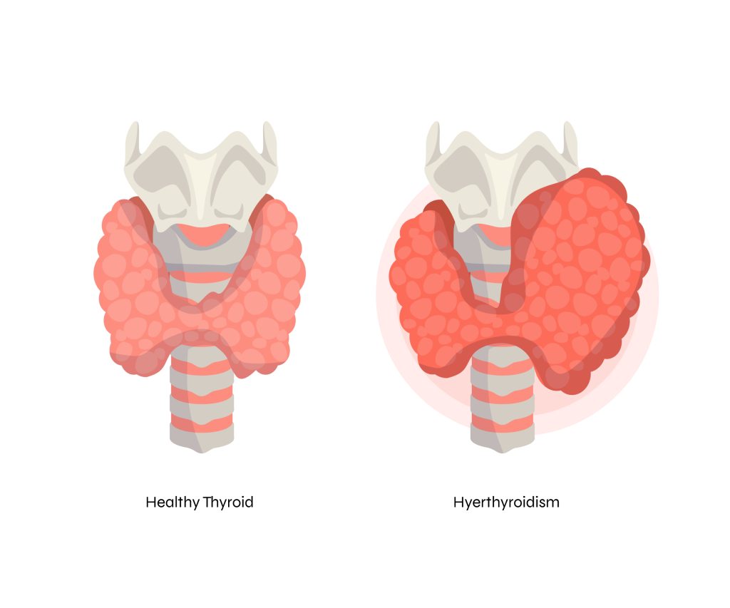
Females are at higher risk of Hypothyroidism Sometimes, newly born babies are also affected with hypothyroidism known as Congenital Hypothyroidism.[One of the most common treatable causes of intellectual disability The thyroid gland weighs about 10 to 20 grams. Lack of iodine causes hypothyroidism and even excess of iodine causes hypothyroidism{Wolff–Chaikoff effect. Table of Contents Overview of THE BUTTERFLY GLAND (The Thyroid) The thyroid is butterfly-shaped structure located in the front of the neck beneath the skin and muscle layer. It’s a part of the endocrine system and controls many of your body’s important functions by producing and releasing (secreting) certain hormones. Secretion of the thyroid hormones T4 (thyroxine) and T3 (triiodothyronine) is regulated by pituitary thyroid-stimulating hormone (TSH). Very small changes in serum-free T4 concentrations induce very large reciprocal changes in serum TSH concentrations. As a result, thyroid function is best assessed by measuring serum TSH, assuming steady-state conditions and the absence of pituitary or hypothalamic disease.  Are you being victim of hypothyroidism? Hypothyroidism accounts for almost 90% cases of thyroid disorder. Do you have these symptoms? {Its time to get your Thyroid Function Test done} How to find out the type of Hypothyroidism correlating with laboratory parameters? [High TSH and low T4] Primary hypothyroidism(almost 95% of cases of hypothyroidism) is characterized by a high serum thyroid-stimulating hormone (TSH) concentration and a low serum free thyroxine (T4) concentration. [High TSH and Normal T4] Subclinical hypothyroidism is defined biochemically as a normal free T4 concentration in the presence of an elevated TSH concentration. {+-TSH and low T4}  Secondary (central) hypothyroidism is characterized by a low serum T4 concentration and a serum TSH concentration that is not appropriately elevated. Is the treatment for hypothyroidism available? Hypothyroidism is corrected with synthetic thyroxine (T4, levothyroxine). Reassess the level or monitoring of TSH should be done after 6 weeks of initiation levothyroxine therapy and adjust the dose accordingly as prescribed by the doctor. Levothyroxine should be taken on an empty stomach so that the body can absorb it fully. Take it at least 1hour  before breakfast. Avoid the use of PPI(Esomeprazole), Iron during Levothyroxine therapy as is interfere its absorption. Credit: UpToDate Research articles.

THYROID DISORDER- An alarming Hypothyroidism? Read More »

Regulation of body temperature 

Body temperature is regulated in the pre optic nucleus of the anterior hypothalamus, which carefully maintains a core temperature of 37°C±1° (98.6˚F±1.8°) A serious threat- Heat Exhaustion and Heat stroke Regulation of body temperature  Body temperature is regulated in the pre optic nucleus of the anterior hypothalamus, which carefully maintains a core temperature of 37°C±1° (98.6˚F±1.8°) Evaporation occurs when water vaporizes from the skin and respiratory tract. This is the body’s most effective mechanism for dissipating excess heat and is the primary means for athletes exercising in hot environments. Radiation is the emission of electromagnetic heat waves. This energy transfer does not require direct contact or air motion. Convection is the transfer of heat to a gas or liquid moving over the body. Heat transfer occurs when the gas or liquid is colder than the body. Conduction is direct heat transfer to an adjacent, cooler object. Heat exhaustion — Heat exhaustion is characterized by the inability to maintain adequate cardiac output due to strenuous physical exercise and environmental heat stress. Most often this manifests as physical collapse during exercise. Body temperature elevation is milder than with EHS or heat injury and the central nervous system is not affected. Risk Factors include: -Sweat with high salt concentration -Heavy sweating -Dehydration -Insufficient sodium intake prior to and during intense activity -Lack of heat acclimatization -Baseline (pre activity) fatigue -History of heat cramps The clinical criteria for heat exhaustion generally include the following: > Athlete has obvious difficulty continuing with exercise. > Core body temperature is usually 101 to 104ºF (38.3 to 40.0ºC) at the time of collapse. > No significant dysfunction of the central nervous system (eg, seizure, altered consciousness, persistent delirium) is present. This is the key difference from exertional heat stroke. Causes of Exhaustion Heat Illness: 1. Strenuous exercise in high ambient temperature and humidity 2. Lack of acclimatization 3. Poor physical fitness 4. Obesity 5. Dehydration 6. Acute illness 7. External load, including clothing, equipment, and protective gear 8. Drugs (Anticholinergic agents),Anti-epileptic agents,Angiotensin-converting enzyme inhibitors,Angiotensin II receptor blockers,Antihistamines,Decongestants Signs and Symptoms of Heat Exhaustion: -Tachycardia and hypotension -Extreme weakness -Dehydration and electrolyte losses -Ataxia and coordination problems, syncope, light-headedness -Profuse sweating, pallor, “prickly heat” sensations -Headache -Abdominal cramps, nausea, vomiting, diarrhea -Persistent muscle cramps How to save the life of patient suffering from Heat Exertion? ~Remove any athlete from play and move them to a shaded or air-conditioned area. ~Place the patient supine with their feet elevated above the level of their head (ie, raise their legs). ~Remove excess clothing and equipment. ~Cool the patient until their rectal temperature is approximately 101ºF (38.3ºC). ~The mode of cooling is less important for heat exhaustion than heat stroke since the cooling is primarily for comfort rather than life-saving. Any technique used to treat heat stroke may be used, including immersing the athlete in a tub of cool water, running cool water over them using a shower or hose, or using evaporative cooling measures. The time needed to reach the goal temperature will be much shorter than with heat stroke. ~Rehydrate the patient with chilled water or a sports drink if they are not nauseated, vomiting, or manifesting a depressed mental status; give IV fluid if the athlete is unable to drink (one reasonable approach is to give a rapid IV bolus of one liter isotonic saline followed by an infusion at a maintenance or 1.5 times maintenance rate, and titrated to response). ~Continuously observe and frequently monitor heart rate, blood pressure, respiratory rate, rectal temperature, and mental status. ~Transport the patient to an emergency department if rapid improvement does not occur despite appropriate treatment Exertional Heat Stroke(EHS) The two main criteria for diagnosing exertional heat stroke (EHS) are 1. A core temperature above 104°F (40°C), measured immediately following collapse during strenuous activity, and CNS dysfunction. CNS dysfunction can manifest as a wide range of possible symptoms and signs, including: disorientation, headache, irrational behavior, irritability, emotional instability, confusion, altered consciousness, coma, or seizure. 2. An athlete should recover within 15 to 20 minutes with these maneuvers; failure to improve should prompt further evaluation, including a rectal temperature. Patients at higher risk for dangerous causes or adverse outcomes and those who do not completely recover within 20 minutes should be evaluated in the emergency department using the approach for any patient with syncope. When the athletes can return back on field after an episode of Heat Related Illness? The recommendations of the American College of Sports Medicine for returning an athlete to training and competition following an episode of EHS represent one reasonable approach : ●No exercise permitted for at least seven days following release from medical care. ●Follow-up with the medical team approximately one week after release for physical examination and any necessary laboratory testing and diagnostic imaging based upon the organs affected during the EHS episode. ●Once cleared for a return to activity, the athlete begins exercise in a cool environment and gradually increases the duration, intensity, and heat exposure over two weeks to demonstrate heat tolerance and initiate acclimatization. ●Athletes who cannot resume vigorous activity over four weeks because of recurrent symptoms (eg, excessive fatigue) should be reevaluated. Laboratory exercise-heat tolerance testing may be useful in this setting. ●The athlete may resume full competition once he or she is able to participate in full training in the heat for two to four weeks without adverse effects.

Regulation of body temperature  Read More »

Interesting facts:

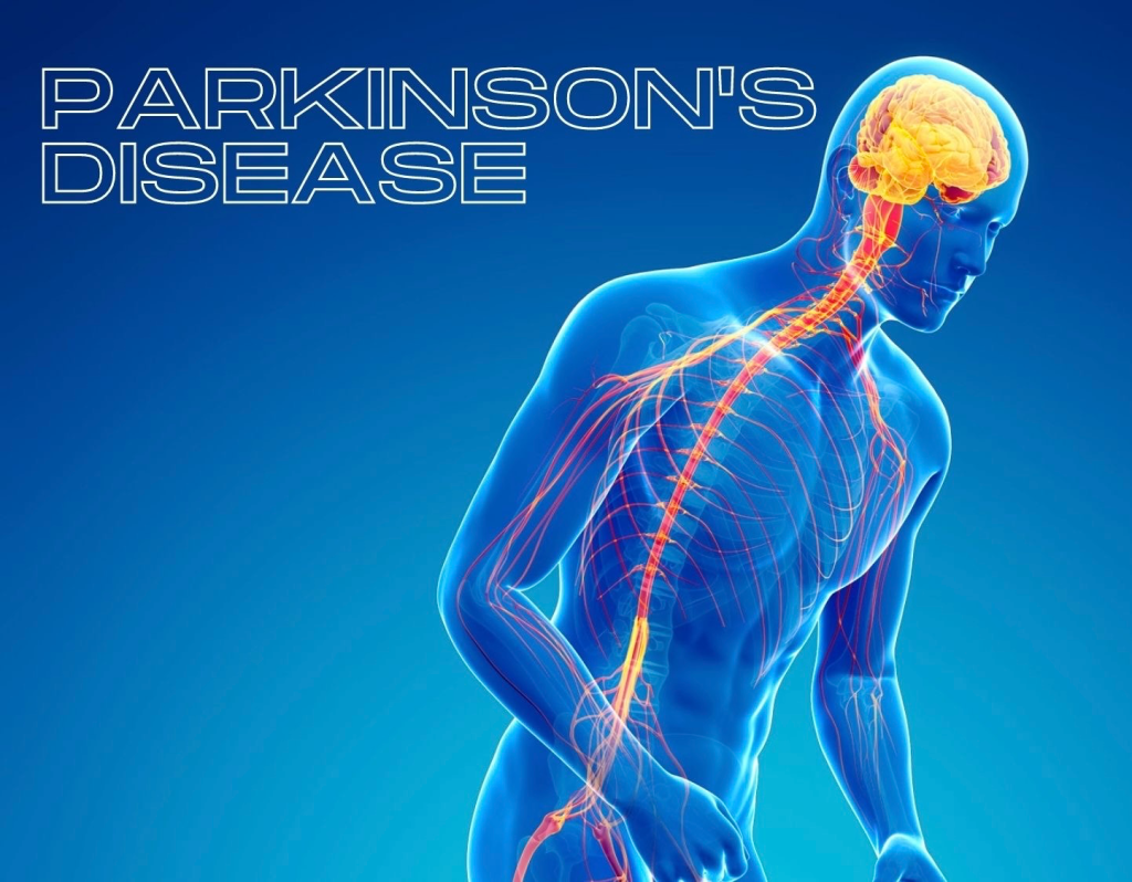
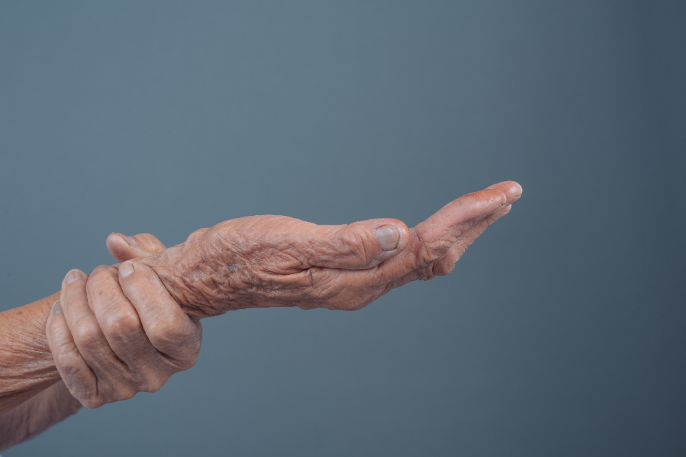
Initially the disease was first described as SHAKING PALSY by James Parkinson. It is usually adult onset Neurodegenerative disease that rises rapidly over the age of 60 years but 10% of the population tend to have the young onset. Risk factors for Parkinson Disease includes: – Age: Beginning of the 5th decade and increases rapidly. – Sex: Male to female ration is 1.4:1 – Genetics – Environmental exposure: Exposure to pesticides, Exposure to air pollution, including nitrogen dioxide and fine particulate matter, High consumption of dairy products, Living in urban or industrial areas with high release of copper, manganese, or lead, Exposure to hydrocarbon solvents, particularly trichloroethylene, Living in rural areas, Farming or agriculture work, The use of well water, High dietary intake of iron, especially in combination with high manganese intake, Reduced levels of dietary and sunlight-derived vitamin D. -Comorbidities includes: Excess body weight and metabolic syndrome, History of prior traumatic brain injury, Type 2 diabetes mellitus, History of melanoma or prostate cancer. Protective factors/Negative associations for the disease includes: -The most consistently identified negative associations, or protective factors, for PD are cigarette smoking, caffeine consumption, and physical exercise. Symptoms that have received the most attention as sensitive prodromal markers ( Initial phase) of PD are: #Rapid eye movement (REM) #Constipation #Hyposmia/olfactory dysfunction #Urinary urgency #Sexual dysfunction #Neuro-cognitive dysfunction. Parkinson Disease is characterized by three main motor features: 1. Tremor: The tremor in PD is a type of rest tremor, meaning that it is most noticeable when the affected body part is supported against gravity and not engaged in purposeful activities. The tremor in the upper extremity is called “pill-rolling” because of the way the thumb and fingers appear to be rolling a small object between them. 2. Bradykinesia: Bradykinesia means generalized slowness of movement but also refers to decreased amplitude of movements. It is also the most difficult symptom for patients to describe. “Weakness,” “incoordination,” and “tiredness” are often used to describe the decreased ability to initiate voluntary movement. 3. Rigidity: Rigidity is an increased resistance to passive movement about a joint. Rigidity can affect any part of the body and may contribute to complaints of stiffness and pain. NONMOTOR SYMPTOMS includes: ●Cognitive dysfunction and dementia ●Psychotic symptoms (hallucinations and delusions) ●Mood disorders including depression, anxiety, and apathy/abulia ●Sleep disturbances ●Fatigue ●Autonomic dysfunction ●Olfactory dysfunction ●Gastrointestinal dysfunction ●Pain and sensory disturbances ●Dermatologic findings

Interesting facts: Read More »

Blood Code

In a world ravaged by the Lost, where the very fabric of reality has been torn asunder, the Revenant rises again, forged from the ashes of a dying world. With each step, the landscape shifts and morphs, like a living, breathing entity, as the Revenant navigates the treacherous terrain. The air is thick with the scent of decay and death, and the only solace lies in the power of the Blood Codes. These ancient artifacts hold the key to unlocking the secrets of the Lost, and the Revenant’s very existence hangs in the balance. The stakes are high, the tension palpable, as the Revenant must confront the darkness within and without. Can they overcome the weight of their own destiny, or will they succumb to the crushing pressure of an unforgiving world? Combat Styles and Supernatural Gifts AI Companions and Online Co-op Deluxe Edition Exclusive Content As the Revenant navigates the ruins of a world gone mad, they must confront the darkness within and without. Will they find a way to reclaim their place in this desolate landscape, or will they succumb to the crushing pressure of an unforgiving world? The choice is theirs, and the fate of the Lost hangs in the balance.

Blood Code Read More »

MILIARIA- SKIN DISORDER

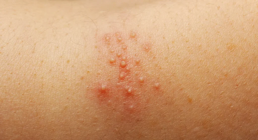
INTRODUCTION “Miliaria” is the general term used to describe conditions caused by blockage within the sweat duct. Eccrine miliaria is a common, transient cutaneous disorder caused by blockage within the eccrine sweat duct. It is also known as “sweat rash,” “prickly heat,” or “heat rash.” CAUSES: Anything that causes sweating can lead to eccrine miliaria in infants, children, and adults. Common causes include: ●Hot and humid environments ●Strenuous physical activity ●Febrile illness [6] ●Occlusion of the skin CLINICAL MANIFESTATIONS The clinical features of miliaria crystallina, miliaria rubra, and miliaria profunda are reviewed below: ●Miliaria crystallina – Results from superficial blockage of the sweat duct (within the stratum corneum) and is characterized by superficial, clear 1 to 2 mm vesicles that resemble water droplets. The vesicles may coalesce. Due to their superficial location, the vesicles rupture easily and do not cause a significant inflammatory response. ●Miliaria rubra – Results from blockage of the duct, usually within the epidermis, although it can occur in the dermis. Leakage of sweat causes inflammation in the periductal tissue. Miliaria rubra is characterized by erythematous 2 to 4 mm papules. They may be papulovesicular or pustular. ●Miliaria profunda – Caused by blockage deeper within the sweat duct (at the dermal-epidermal junction or below). It usually occurs after repeated episodes of miliaria rubra. Escape of sweat into the surrounding tissues results in erythematous to skin-colored, firm papules 1 to 4 mm in diameter. The papules are non-follicular. It is most common in adult males, especially military personnel stationed in tropical climates Seen most in adults, miliaria profunda usually occurs on the trunk but can be seen on the extremities as well. Because sweat is obstructed deeply within the skin, affected areas show little or no sweating. The eruption is typically asymptomatic. It may also be subtle and become more visible when the patient sweats. ●Management •General measures – Treatment of miliaria revolves around minimizing exposure to factors that stimulate sweating and occlusion of eccrine sweat glands. This approach includes the following measures: -Move patient to a cooler environment if possible -Wear breathable clothing (such as cotton) that does not occlude the skin -Remove occlusive bandages or medication patches in the affected area and use more porous alternatives if needed -Treat fever with antipyretics

MILIARIA- SKIN DISORDER Read More »

Treatment of Malaria

For Uncomplicated Falciparum malaria (Adult >35 kg) The first line treatment for falciparum malaria is artemether + lumefantrine (AL) given over three days and a single dose primaquine.  (6 doses) STAT-8Hrs- then BD for 2 more days. NOTE: Take after a full meal or whole milk. If a patient vomits within 30 minutes of taking a dose, he or she should repeat the dose. Ideally, the first two doses should be taken 8 hours apart. For Complicated Falciparum Malaria (Severe): Intravenous or intramuscular artesunate for at least 24 h and until the patient can tolerate oral medication. Once a patient has received at least 24h of parenteral therapy and can tolerate oral therapy, complete treatment with full course artemether + lumefantrine (AL). Inj. Artesunate 2.4 mg/kg STAT, 12 hours and 24hours and then OD until oral medication is tolerated. If Artemisinin combination therapy is not available and chloroquine sensitive species. Dosage Distribution: Chloroquine- Day 1: chloroquine is given at an initial dose of 10 mg base/kg body weight, Day 2: followed by 10 mg/kg body weight, Day 3: 5 mg/kg body weight. Primaquine- 0.25 mg/kg body weight per day. Artemether and Lumefantrine- Total dose of 5-24 mg/kg of artemether and 29-144 mg /kg of lumefantrine. Malaria Types of species that cause malaria: What causes malaria? High risk individuals for malaria Symptoms of Malaria: Clinical findings include: 

How does malaria get transmitted? Following the bite of an infected female Anopheles mosquito, the inoculated sporozoites migrate to the liver within one to two hours. Individuals are generally asymptomatic for 12 to 35 days after infection, but symptoms can commence as early as 7 days (depending on parasite species). Symptoms begin during the erythrocytic stage of the parasite life cycle, when infected red cells rupture and release merozoites, leading to fever and other symptoms. In most cases, infections due to P. falciparum become clinically apparent within one month after exposure. Longer incubation periods are more likely in semi-immune individuals and individuals taking incompletely effective malaria prophylaxis. The incubation period for the relapsing species, Plasmodium vivax and Plasmodium ovale, is also about two weeks; however, illness can occur months after initial infection due to activation of residual hypnozoites in the liver. Relapses generally occur within two to three years of infection; with even longer periods of dormancy being reported. The incubation period for Plasmodium malariae is about 18 days; however, low-grade asymptomatic infections can very rarely persist for years. P. falciparum and P. malariae have no dormant (hypnozoite) phase, hence do not relapse.

Treatment for Malaria: The treatment depends on the species of plasmodium.
-Symptomatic management includes Paracetamol -Oral medication includes: Artemether, Lumefantrine, Chloroquine, Primaquine -Injectable medication includes: INJ Artesunate For Uncomplicated Falciparum malaria (Adult >35 kg) The first line treatment for falciparum malaria is artemether + lumefantrine (AL) given over three days and a single dose primaquine.  (6 doses) STAT-8Hrs- then BD for 2 more days. NOTE: Take after a full meal or whole milk. If a patient vomits within 30 minutes of taking a dose, he or she should repeat the dose. Ideally, the first two doses should be taken 8 hours apart. For Complicated Falciparum Malaria (Severe): Intravenous or intramuscular artesunate for at least 24 h and until the patient can tolerate oral medication. Once a patient has received at least 24h of parenteral therapy and can tolerate oral therapy, complete treatment with full course artemether + lumefantrine (AL). Inj. Artesunate 2.4 mg/kg STAT, 12 hours and 24hours and then OD until oral medication is tolerated. If Artemisinin combination therapy is not available and chloroquine sensitive species. Dosage Distribution: Chloroquine- Day 1: chloroquine is given at an initial dose of 10 mg base/kg body weight, Day 2: followed by 10 mg/kg body weight, Day 3: 5 mg/kg body weight. Primaquine- 0.25 mg/kg body weight per day. Artemether and Lumefantrine- Total dose of 5-24 mg/kg of artemether and 29-144 mg /kg of lumefantrine. Malaria Types of species that cause malaria: What causes malaria? High risk individuals for malaria Symptoms of Malaria: Clinical findings include: 

How does malaria get transmitted? Following the bite of an infected female Anopheles mosquito, the inoculated sporozoites migrate to the liver within one to two hours. Individuals are generally asymptomatic for 12 to 35 days after infection, but symptoms can commence as early as 7 days (depending on parasite species). Symptoms begin during the erythrocytic stage of the parasite life cycle, when infected red cells rupture and release merozoites, leading to fever and other symptoms. In most cases, infections due to P. falciparum become clinically apparent within one month after exposure. Longer incubation periods are more likely in semi-immune individuals and individuals taking incompletely effective malaria prophylaxis. The incubation period for the relapsing species, Plasmodium vivax and Plasmodium ovale, is also about two weeks; however, illness can occur months after initial infection due to activation of residual hypnozoites in the liver. Relapses generally occur within two to three years of infection; with even longer periods of dormancy being reported. The incubation period for Plasmodium malariae is about 18 days; however, low-grade asymptomatic infections can very rarely persist for years. P. falciparum and P. malariae have no dormant (hypnozoite) phase, hence do not relapse.

Treatment for Malaria: The treatment depends on the species of plasmodium.
-Symptomatic management includes Paracetamol -Oral medication includes: Artemether, Lumefantrine, Chloroquine, Primaquine -Injectable medication includes: INJ Artesunate Severe Malaria

Treatment of Malaria Read More »